43 cell diagram and labels
CELL MEMBRANE LABEL Diagram | Quizlet identifies or labels the cell. Receptor protein. receives information. Heads. part of the phospholipid that loves water (hydrophili) - points to the most outside and inside of cell. Tails. part of phospholipid that hates water (hydrophobic); points to the interior or Inside. Phospholipid Bilayer. 2 layers of fat - tails point in toward each ... Animal Cell Diagram with Label and Explanation: Cell ... - Collegedunia Diagram of Animal Cell Below is the diagram of the animal cell which shows the organelles present in it. The cell is covered with cytoplasm which consists of cell organelles in it. The nucleus is covered with a rough Endoplasmic Reticulum and other organelles each designed for a specific purpose.
Interactive Cell Cycle - CELLS alive INTERPHASE. Gap 0. Gap 1. S Phase. Gap 2. MITOSIS . ^ Cell Cycle Overview Cell Cycle Mitosis > Meiosis > Get the Cell Division PowerPoints
Cell diagram and labels
A Well-labelled Diagram Of Animal Cell With Explanation - BYJUS The animal cell diagram is widely asked in Class 10 and 12 examinations and is beneficial to understand the structure and functions of an animal. A brief explanation of the different parts of an animal cell along with a well-labelled diagram is mentioned below for reference. Also Read Different between Plant Cell and Animal Cell Animal Cells: Labelled Diagram, Definitions, and Structure - Research Tweet The endoplasmic reticulum (s) are organelles that create a network of membranes that transport substances around the cell. They have phospholipid bilayers. There are two types of ER: the rough ER, and the smooth ER. The rough endoplasmic reticulum is rough because it has ribosomes (which is explained below) attached to it. Skin Diagram with Detailed Illustrations and Clear Labels - BYJUS Explore Skin Diagram with BYJU’S. Diagram of the skin is illustrated in detail with neat and clear labelling. Also available for free download. Login. Study Materials. ... Human Cell Structure: Types Of Microbes: Biome Meaning: What Is A Neuron: 1 Comment. Neeraj Shukla September 23, 2021 at 1:28 pm. Up board English medium. Reply.
Cell diagram and labels. Nuclear envelope - Wikipedia The nuclear envelope is punctured by around a thousand nuclear pore complexes, about 100 nm across, with an inner channel about 40 nm wide. The complexes contain a number of nucleoporins, proteins that link the inner and outer nuclear membranes.. Cell division. During the G2 phase of interphase, the nuclear membrane increases its surface area and doubles its … Animal cells and plant cells - Cells to systems - BBC Bitesize Part Function Found in; Cell membrane: Controls the movement of substances into and out of the cell: Plant and animal cells: Cytoplasm: Jelly-like substance, where chemical reactions happen 8.6.1.1 Adding Unicode and ANSI Characters in Text Labels - Origin Adding Unicode Characters to Text Labels. There are various ways to add Unicode characters to your text labels. While creating your text label, enter the 4-character hex code for the character (e.g. 03BB for "λ"), then press ALT+X.; Versions prior to 2022b: While creating your text label, right-click and choose Symbol Map.Select your Font, check the Unicode box and enter the 4 … Labeled Plant Cell With Diagrams | Science Trends The parts of a plant cell include the cell wall, the cell membrane, the cytoskeleton or cytoplasm, the nucleus, the Golgi body, the mitochondria, the peroxisome's, the vacuoles, ribosomes, and the endoplasmic reticulum. Parts Of A Plant Cell The Cell Wall Let's start from the outside and work our way inwards.
Cell: Structure and Functions (With Diagram) - Biology Discussion Eukaryotic Cells: 1. Eukaryotes are sophisticated cells with a well defined nucleus and cell organelles. 2. The cells are comparatively larger in size (10-100 μm). 3. Unicellular to multicellular in nature and evolved ~1 billion years ago. 4. The cell membrane is semipermeable and flexible. 5. These cells reproduce both asexually and sexually. A Labeled Diagram of the Animal Cell and its Organelles A Labeled Diagram of the Animal Cell and its Organelles There are two types of cells - Prokaryotic and Eucaryotic. Eukaryotic cells are larger, more complex, and have evolved more recently than prokaryotes. Where, prokaryotes are just bacteria and archaea, eukaryotes are literally everything else. Human Cell Diagram, Parts, Pictures, Structure and Functions One of the few cells in the human body that lacks almost all organelles are the red blood cells. The main organelles are as follows : cell membrane endoplasmic reticulum Golgi apparatus lysosomes mitochondria nucleus perioxisomes microfilaments and microtubules Diagram of the human cell illustrating the different parts of the cell. Cell Membrane Labeling a Cell Diagram | Quizlet Cell Wall This gives shape and support to the plant cell. It surrounds the cell and protects the other parts of the cell. Chloroplasts This is where the plant cell's chlorophyll is stored. This is what the plant uses to make its own food (photosynthesis). This is also what makes plant cells have a green-like color. Plant cells Are circular in shape
Converting Diagrams - The Biology Corner Open Google Draw and import the diagram. Then use "insert" to create text boxes where students can fill in the labels. Don't forget when assigning this to students on Google classroom to make a copy for each student. You can leave documents in an uneditable form and students can use an addon like "Kami" to annotate the document. Plant Cell - Definition, Structure, Function, Diagram & Types - BYJUS The primary function of the cell wall is to protect and provide structural support to the cell. The plant cell wall is also involved in protecting the cell against mechanical stress and providing form and structure to the cell. It also filters the molecules passing in and out of it. The formation of the cell wall is guided by microtubules. Plant Cell: Diagram, Types and Functions - Embibe Exams Q.2. How to make a model of a plant cell diagram step by step procedure? Ans: The plant cell diagram can be checked above and on a similar pattern the diagram can be created. Q.3. Why do plant cells possess large-sized vacuoles? Ans: Vacuole functions in the storage of substances, maintenance of osmolarity and sustaining turgor pressure. Q.4. Animal Cell - Structure, Function, Diagram and Types - BYJUS The cell is the structural and functional unit of life. These cells differ in their shapes, sizes and their structure as they have to fulfil specific functions. Plant cells and animal cells share some common features as both are eukaryotic cells. However, they differ as animals need to adapt to a more active and non-sedentary lifestyle.
Cell Diagram | Free Cell Diagram Templates - Edrawsoft A free customizable cells diagram template is provided to download and print. Quickly get a head-start when creating your own cell diagram. Here is a simple cell diagram example created by Science Diagram Maker Download Template: Get EdrawMax Now! Free Download Popular Latest Flowchart Process Flowchart Workflow BPMN Cross-Functional Flowchart
How to plot a ternary diagram in Excel - Chemostratigraphy.com Feb 13, 2022 · Adding labels to the apices. Next, we need some space for the apices labels: click into the Plot Area (not the Chart Area) then resize by holding the Shift key (this ensures an equal scaling) and use the mouse cursor on one of the corner pick-points. Then recentre the Plot Area in the Chart Area.

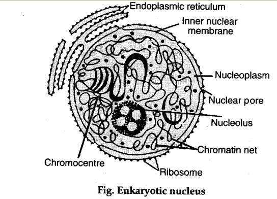
Describe the structure of nucleus and centrosome with the help of labelled diagram - CBSE Class ...
How to Create Venn Diagram in Excel – Free Template Download Replace the default values with the custom labels you previously designed. Right-click on any data label and choose “Format Data Labels.” Once the task pane pops up, do the following: Go to the Label Options tab. Click “Value From Cells.” Highlight the corresponding cell from column Label (H2 for Coca-Cola, H3 for Pepsi, and H4 for Dr ...
Essential JS 2 for Angular - Syncfusion Essential JS 2 for Angular is a modern JavaScript UI toolkit that has been built from the ground up to be lightweight, responsive, modular and touch friendly. It is written in TypeScript and has no external dependencies.
quiver: a modern commutative diagram editor Welcome. quiver is a modern, graphical editor for commutative and pasting diagrams, capable of rendering high-quality diagrams for screen viewing, and exporting to LaTeX via tikz-cd.. quiver is intended to be intuitive to use and easy to pick up. Here are a few tips to help you get started: Click and drag to create new arrows: the source and target objects will be created automatically.
Free Cell Diagram Software with Free Templates - EdrawMax - Edrawsoft An animal cell diagram describes a cell structure enclosed by a plasma member, and it has a nucleus with a membrane and organelles. Neuron Diagram A neuron diagram describes the three parts of a Neuron: dendrites, an axon, a cell body, or soma. Cell Membrane Diagram
PDF Human Cell Diagram, Parts, Pictures, Structure and Functions One of the few cells in the human body that lacks almost all organelles are the red blood cells. The main organelles are as follows : cell membrane endoplasmic reticulum Golgi apparatus lysosomes mitochondria nucleus perioxisomes microfilaments and microtubules 2
2,141 Red blood cell diagram Images, Stock Photos & Vectors | Shutterstock Find Red blood cell diagram stock images in HD and millions of other royalty-free stock photos, illustrations and vectors in the Shutterstock collection. Thousands of new, high-quality pictures added every day.
Learn the parts of a cell with diagrams and cell quizzes Cell diagram unlabeled It's time to label the cell yourself! As you fill in the cell structure worksheet, remember the functions of each part of the cell that you learned in the video. Doing this will help you to remember where each part is located. Click the links below to download the labeled and unlabeled eukaryotic cell diagrams.
Interactive Cell Model - CELLS alive Cell Wall. Chloroplast. Smooth Endoplasmic Reticulum. Rough Endoplasmic Reticulum. Ribosomes. Cytoskeleton. RETURN to CELL DIAGRAM ...
03 Label the Cell Diagram | Quizlet Start studying 03 Label the Cell. Learn vocabulary, terms, and more with flashcards, games, and other study tools.
A Labelled Diagram Of Neuron with Detailed Explanations - BYJUS A neuron is a specialized cell, primarily involved in transmitting information through electrical and chemical signals. They are found in the brain, spinal cord and the peripheral nerves. ... Diagram Of Neuron with Labels. Here is the description of human neuron along with the diagram of the neuron and their parts.
Skin Diagram with Detailed Illustrations and Clear Labels - BYJUS Explore Skin Diagram with BYJU’S. Diagram of the skin is illustrated in detail with neat and clear labelling. Also available for free download. Login. Study Materials. ... Human Cell Structure: Types Of Microbes: Biome Meaning: What Is A Neuron: 1 Comment. Neeraj Shukla September 23, 2021 at 1:28 pm. Up board English medium. Reply.
Animal Cells: Labelled Diagram, Definitions, and Structure - Research Tweet The endoplasmic reticulum (s) are organelles that create a network of membranes that transport substances around the cell. They have phospholipid bilayers. There are two types of ER: the rough ER, and the smooth ER. The rough endoplasmic reticulum is rough because it has ribosomes (which is explained below) attached to it.
A Well-labelled Diagram Of Animal Cell With Explanation - BYJUS The animal cell diagram is widely asked in Class 10 and 12 examinations and is beneficial to understand the structure and functions of an animal. A brief explanation of the different parts of an animal cell along with a well-labelled diagram is mentioned below for reference. Also Read Different between Plant Cell and Animal Cell


/plant_anaphase-56a09b0d3df78cafdaa32db4.jpg)


/2000px-Plant_cell_structure_svg_vacuole.svg-58a886443df78c345bf8d009.png)

Post a Comment for "43 cell diagram and labels"