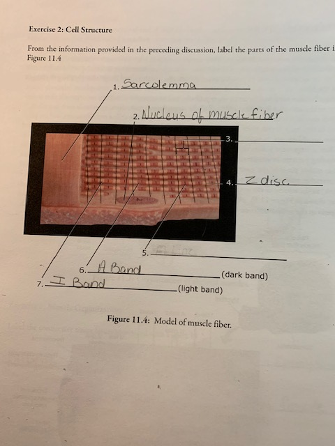
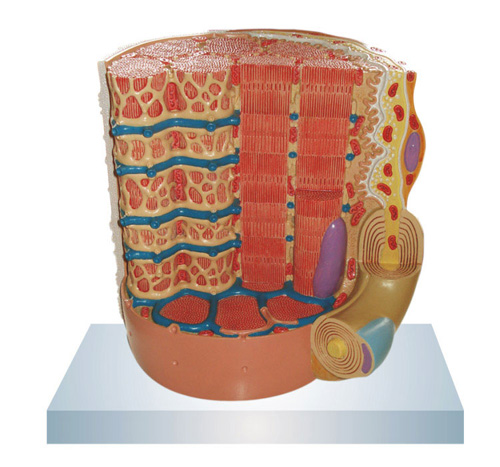
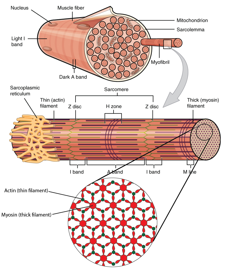
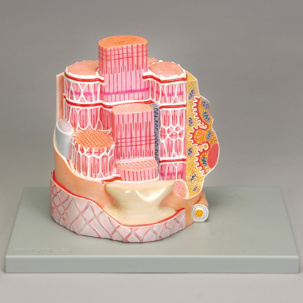
38 muscle fiber model with labels
muscle fiber model labeling Diagram | Quizlet Start studying muscle fiber model labeling. Learn vocabulary, terms, and more with flashcards, games, and other study tools. Andrew File System Retirement - Technology at MSU Andrew File System (AFS) ended service on January 1, 2021. AFS was a file system and sharing platform that allowed users to access and distribute stored content. AFS was available at afs.msu.edu an…
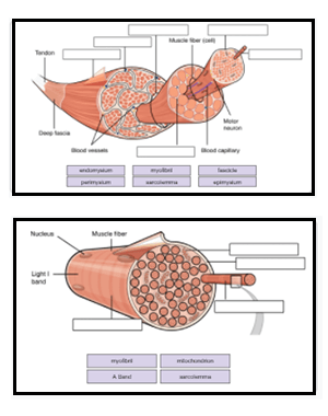
20 Unlabeled Muscle Diagram Worksheet | Worksheet From Home 20 Unlabeled Muscle Diagram Worksheet. Label Muscles Worksheet unlabeled male reproductive system, unlabeled muscle fiber, unlabeled muscles, unlabeled muscular system image, unlabeled male reproductive system diagram, via: pinterest.com. Numbering Worksheets for Kids. Kids are usually introduced to this topic matter during their math education.
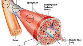
Muscle fiber model with labels
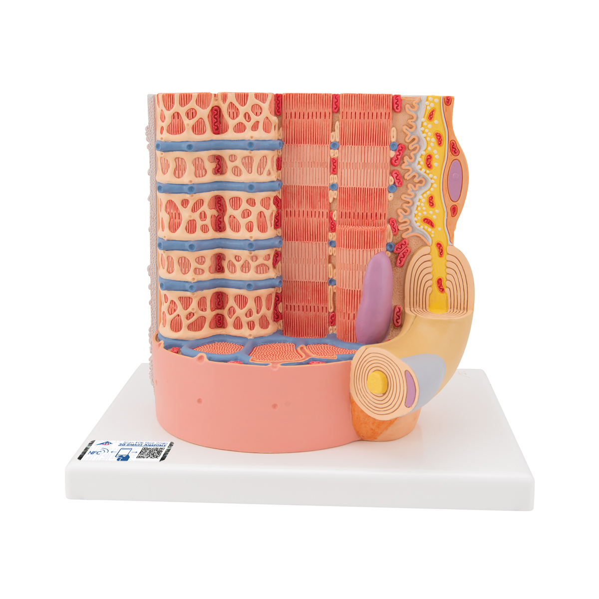
muscle fiber diagram labeled muscle muscles anterior body human organs diagram anatomy gray system muscular musculoskeletal wikidoc facts basic skeletal major diagrams weight types. Neuromuscular Junction Model Labeled - Google Search | Anatomy . junction neuromuscular anatomy muscle labeled skeletal sarcomere physiology system google fiber muscular ... Skeletal muscle tissue: Histology | Kenhub Type I muscle fibers, also called slow oxidative fibers, are specialized for aerobic activity. They are small, contain a high amount of myoglobin, and appear red in fresh tissue. ... Try our tissue quizzes and free labeling worksheets! Sarcomeres. The sarcomere is the functional unit of a skeletal muscle cell. Each sarcomere is about 2.5 ... Muscle Models | Muscle Figures | Musculature Models - 3B Scientific 3B MICROanatomy™ Human Muscle Fiber Model, 10,000 times magnified - 3B Smart Anatomy $ 339.00 Item: 1000213 [B60] This micro-anatomy model magnifies the anatomy of the human muscle fiber approximately 10,000 times. This muscle model illustrates a section of a skeletal muscle fiber and its neuromuscular end plate.
Muscle fiber model with labels. Muscle Fiber Model (Altay) Flashcards | Quizlet Muscle fiber model identifications Terms in this set (21) sarcolemma Identify the membrane. endomysium Identify the tissue layer. myofibril Identify the structure. thick myofilament Identify the structure. thin myofilament Identify the structure. neuromuscular junction Identify the connection. axon Identify the structure. axon terminals Solved Lab 9: Muscle Tissue and axial muscle Exercise 1. | Chegg.com Identify and list a function of each labeled item 1-8) on the models Below. These are the same model at different angles. These are models of ONE muscle fiber or one skeletal muscle cell. Use the following terms to help you label: Endomysium, sarcolemma, myofibril, sarcoplasmic reticulum, myofilaments, motor neuron, T-tubule, nucleus. Trigger Point Therapy – That Is How We Treat Pain Jan 26, 2017 · a taut band (muscle fiber bundle) in the muscle; a pressure-sensitive area within the taut band; referred pain from a trigger point; a local twitch response of the trigger point or taut band in response to mechanical stimulation of the trigger point. These diagnostic criteria have been shown to have a high intertester reliability in trained ... Amazon.com: Muscle Milk Genuine Protein Shake, Strawberries … About this item . Contains twelve (12) 11 fl oz Cartons of Muscle Milk Genuine Protein Shakes. Packaging may vary. HELPS SATISFY HUNGER AND BUILD MUSCLE – Muscle Milk Genuine is an energizing protein shake that can be consumed as an on-the-go breakfast or anytime snack or to support post-workout recovery and muscle growth.
muscle man labeled anatomy 3d Athletic Male Human Anatomy And Muscles Stock Photo - Image: 29069070. 9 Images about Athletic Male Human Anatomy And Muscles Stock Photo - Image: 29069070 : Athletic Male Human Anatomy And Muscles Stock Photo - Image: 29069070, ᐈ Back muscle diagrams labeled stock vectors, Royalty Free trapezius and also Labeled Skin Model 3D - HAIR FOLLICLE RETICULAR DERMIS ECCRINE SWEAT. Altay Skeletal Muscle Fiber Model | Carolina.com Altay®. 10,000× life size. Microstructure of a skeletal muscle fiber is represented in great detail; includes the neuromuscular junction, sarcoplasmic reticulum, T tubules, and myofibrils. Mounted on reinforced polymer base. Size, 25 × 23 × 23 cm. Skeletal Muscle Fiber Model - Myofibrils - YouTube This video was produced to help students of human anatomy at Modesto Junior College study our anatomical models. Anatomical Models and Keys: Anatomy Models - NEOMED Saunders Heart Model . Female Pelvis on Stand. Denoyer Geppert Heart. Kidney Microanatomy . Stomach. Muscle Fiber. Bone Structure. Liver Microanatomy . Heart and Diaphragm . Larynx . Base of Head . Small Liver Model . Liver Denoyer Model . Kidney Denoyer Model Disc/MRI Head . Smooth Muscle . Heart and Lung Larynx is missing . Brain Stem ...
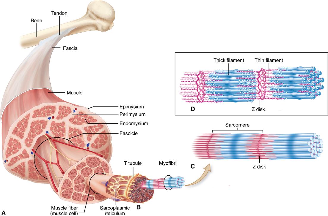
Muscle Fibers: Anatomy, Function, and More - Healthline Each muscle fiber contains smaller units made up of repeating thick and thin filaments. This causes the muscle tissue to be striated, or have a striped appearance. Skeletal muscle fibers are... Muscle Fiber Anatomy Quiz - PurposeGames.com This is an online quiz called Muscle Fiber Anatomy. There is a printable worksheet available for download here so you can take the quiz with pen and paper. Your Skills & Rank. Total Points. 0. Get started! Today's Rank--0. Today 's Points. One of us! Game Points. 15. You need to get 100% to score the 15 points available. Muscle Fiber Model #1 - Ohio University - Anatomy & Physiology Muscle Fiber Model #1 - Ohio University - Anatomy & Physiology SAC A&P Model Key - Muscular System - austincc.edu Muscular System. M1 - Muscled Arm. M2 - Muscle Leg. M3 - Female Muscle Figure. M4 - Microanatomy Muscle Fiber. M5 - Muscle Figure.
Muscle Fiber Model: Motor Neuron, Myeline Sheath, Node of Ranvier ... Muscle Fiber Model: Motor Neuron, Myeline Sheath, Node of Ranvier, Synaptic Terminal, Synaptic Cleft, Endomysium, Sarcolemma, Nuclei, Mitochondria, T-tubules, Sarcoplasmic Reticulum, Myofibrils Find this Pin and more on Strength Training Benefits by Great Bones. Motor Neuron Cell Model Musculoskeletal System Anatomy Models Muscular System
Muscle Cell (Myocyte): Definition, Function & Structure | Biology A single muscle cell contains many nuclei, which are pressed against the cell membrane. A muscle cell is a long cell compared to other forms of cells, and many muscle cells connect together to form the long fibers found in muscle tissue. Structure of a Muscle Cell. As seen in the image below, a muscle cell is a compact bundle of many myofibrils.
anatomy of muscle structure labeling muscle fiber anatomy cell human neuron synaptic body muscles motor models labeled skeletal muscular ranvier mitochondria cleft sarcolemma node skin Canaliculi osteon anatomy physiology bis. Crayfish dissection anatomy labeled external cephalothorax abdomen structure appendages guide science head function parts side segments main label tail region.
Muscle Fiber Labeling Quiz - PurposeGames.com About this Quiz This is an online quiz called Muscle Fiber Labeling Quiz There is a printable worksheet available for download here so you can take the quiz with pen and paper. Your Skills & Rank Total Points 0 Get started! Today's Rank -- 0 Today 's Points One of us! Game Points 17 You need to get 100% to score the 17 points available Actions
Muscle Fiber. 1. Myofibrils 2. Mitochondrium 3 ... - Pinterest Muscle Fiber. 1. Myofibrils 2. Mitochondrium 3. Postsynaptic membrane 4. Synaptic gap with basal lamina 5. Presynaptic membrane 6. Presynaptic vesicle 7. Schwann cell 8. Nucleus 9. Actin filament 10. Sarcomere 11. Myosin filament 12. Myelin sheath 13. Neurofibers 14. Cell membrane (sarcolemma) 15. Transverse membrane tube 16. Triad 17.
muscle anatomy labeling arm anatomy muscle muscles human hand body reference. Skeletal Muscle Fiber Model Quiz . muscle skeletal fiber quiz games purposegames. Label The White Rat Muscles . rat muscles purposegames. Kidney labeled structure human anatomy label labels function labeling systems body final. Muscles of the hand.
Sarcomere Model | Muscle Fiber Model | Skeletal Muscle | MICROanatomy ... This micro-anatomy model magnifies the anatomy of the human muscle fiber approximately 10,000 times. This muscle model illustrates a section of a skeletal muscle fiber and its neuromuscular end plate. The muscle fiber is the basic element of the diagonally striped skeletal muscle. You've never seen a muscle fiber in this way!
Skeletal Muscle Fiber - GetBodySmart Skeletal Muscle Fiber. Skeletal muscles are the types of muscle tissue that enable us with voluntary movements. They are attached to the bones of the skeleton by tendons. Skeletal muscle fibers have a striated (striped) appearance on histological sections because they are made up of smaller units called sarcomeres that run parallel to each other, giving the muscle the striated appearance.
Anabolic steroid - Wikipedia Anabolic steroids, also known more properly as anabolic–androgenic steroids (AAS), are steroidal androgens that include natural androgens like testosterone as well as synthetic androgens that are structurally related and have similar effects to testosterone. They increase protein within cells, especially in skeletal muscles, and also have varying degrees of virilizing effects, including ...
Skeletal Muscle Histology Slide Identification and Labeled Diagram ... Please try to find out these structures from the skeletal muscle slide labeled images. #1. Longitudinal section of skeletal muscle #2. Cross-section of skeletal muscle #3. Skeletal muscle fibers of the longitudinal section #3. The nucleus of skeletal muscle fibers in longitudinal and cross-section #4. Cross striations of skeletal muscles #5.
interactive muscle labeling muscle junction neuromuscular fiber anatomy muscular system. Anatomy Games - Real Bodywork . anatomy muscles game muscle games labeling body label matching human interactive learn major learning labels names upper bodywork alive realbodywork. Anatomy Matching Game - Anatomy Drawing Diagram sen842cova.blogspot.com. nurses. 32 ...
Ultrastructure of Muscle - Skeletal - Sliding Filament - TeachMeAnatomy The sliding filament model describes the mechanism of skeletal muscle contraction Actin and Myosin Muscle fibres are formed from two contractile proteins - actin and myosin. Myosin filaments have many heads, which can bind to sites on the actin filament. Actin filaments are associated with two other regulatory proteins, troponin and tropomyosin.
Learn all muscles with quizzes and labeled diagrams | Kenhub Labeled diagram View the muscles of the upper and lower extremity in the diagrams below. Use the location, shape and surrounding structures to help you memorize each muscle. Once you're feeling confident, it's time to test yourself. Unlabeled diagram See if you can label the muscles yourself on the worksheet available for download below.
Muscles Labeling - The Biology Corner The activity linked below is a drag and drop activity for students to practice labeling the muscles, there are 6 slides showing images of muscles and fibers and the connective tissue surrounding the fibers (endomysium, perimysium, epimysium). Google Slides Key (TpT) Prev Article Next Article
PDF Anatomy & Physiology - TMCC Subcutis (Hypodermis) 1. External Horny Layer (Stratum corneum) 1a. Clear Layer (Stratum lucidum) -(KS 3 only) 2. Internal Hornless Germinative Zone (Stratum germinativum) 2a. Granular Layer (Stratum granulosum) 2b. Prickle-cell Layer (Stratum spinosum) 2c. Cylindrical Layer (Stratum basale) 3. Papillae 4.
Muscle Models | Muscle Figures | Musculature Models - 3B Scientific 3B MICROanatomy™ Human Muscle Fiber Model, 10,000 times magnified - 3B Smart Anatomy $ 339.00 Item: 1000213 [B60] This micro-anatomy model magnifies the anatomy of the human muscle fiber approximately 10,000 times. This muscle model illustrates a section of a skeletal muscle fiber and its neuromuscular end plate.
Skeletal muscle tissue: Histology | Kenhub Type I muscle fibers, also called slow oxidative fibers, are specialized for aerobic activity. They are small, contain a high amount of myoglobin, and appear red in fresh tissue. ... Try our tissue quizzes and free labeling worksheets! Sarcomeres. The sarcomere is the functional unit of a skeletal muscle cell. Each sarcomere is about 2.5 ...
muscle fiber diagram labeled muscle muscles anterior body human organs diagram anatomy gray system muscular musculoskeletal wikidoc facts basic skeletal major diagrams weight types. Neuromuscular Junction Model Labeled - Google Search | Anatomy . junction neuromuscular anatomy muscle labeled skeletal sarcomere physiology system google fiber muscular ...


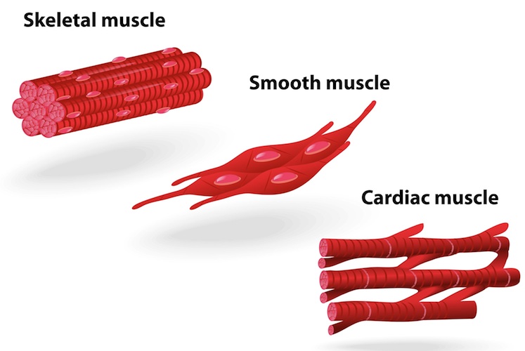

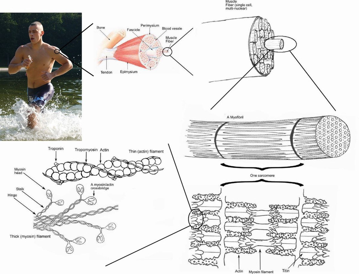
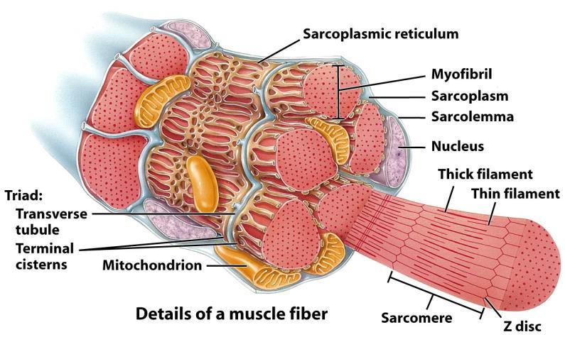











Post a Comment for "38 muscle fiber model with labels"