43 cell wall diagram with labels
Labeled Plant Cell With Diagrams - Science Trends The parts of a plant cell include the cell wall, the cell membrane, the cytoskeleton or cytoplasm, the nucleus, the Golgi body, the mitochondria, the peroxisome's, the vacuoles, ribosomes, and the endoplasmic reticulum. Parts Of A Plant Cell The Cell Wall Let's start from the outside and work our way inwards. › basic-cells › label-diagramLabel Cell Parts | Plant & Animal Cell Activity | StoryboardThat Have your students label a plant and animal cell using one of the landscape poster layouts (small or large). Students will create a cell diagram labeled with the different organelles of plant and animal cells. The cell diagrams are easily colorable, allowing students to differentiate the different parts of the plant and animal cell quickly.
owlcation.com › stem › 3d-cell-modelHow to Create 3D Plant Cell and Animal Cell Models for ... Sep 10, 2011 · Step 1: Choose Plant Cell vs. Animal Cell. First and foremost, you need to decide whether you will create a plant cell or animal cell. Plant cells and animal cells are shaped differently and contain different parts.
Cell wall diagram with labels
flowingdata.com › 2012/10/09 › history-of-History of Earth in 24-hour clock - FlowingData Oct 09, 2012 · I’m sorry but this infographic could have been better server is a different format. I’m really not a fan of this clock. First it’s confusing because it’s a 24 hour clock but the times are given in military time e.g. Sexual Reproduction is at 16:08. Structure of Fungal Cell (With Diagram) | Fungi In this article we will discuss about the structure of fungal cell. This will also help you to draw the structure and diagram of the fungal cell. (a) The Cell Wall of the Fungal Cell: The composition of cell wall is variable among the different groups of fungi or between the different species of the same group. Cell Wall - Definition, Cell Wall Function, Cell Wall Layers A cell wall is defined as the non-living component, covering the outmost layer of a cell. Its composition varies according to the organism and is permeable in nature. The cell wall separates the interior contents of the cell from the exterior environment. It also provides shape, support, and protection to the cell and its organelles.
Cell wall diagram with labels. scioly.org › wiki › indexCell Biology - Wiki - Scioly.org Oct 08, 2021 · Cell membrane: Functions in transport, the movement of substances in and out of the cell, and in energy production (breakdown of large molecules, photosynthesis). Cell wall: Gives structural strength (rigidity) to the cell. Capsule or slime layer: Jelly-like substance which protects the cell wall from environmental damage. Nucleoid A Well-labelled Diagram Of Animal Cell With Explanation The animal cell diagram is widely asked in Class 10 and 12 examinations and is beneficial to understand the structure and functions of an animal. A brief explanation of the different parts of an animal cell along with a well-labelled diagram is mentioned below for reference. Also Read Different between Plant Cell and Animal Cell Structure of Cell Wall (With Diagram) | Plants Cells with secondary wall consist of five layers a three layered secondary wall, the primary wall and the middle lamella. In some cells, such as primary xylem, the secondary thickening materials are laid down in such a way that various patterns are formed on the cell wall, e.g. annular, spiral, reticulate, scalariform and pitted. Structure of Bacterial Cell (With Diagram) These are long filamentous, cytoplasmic appendages, 12-30 μm in length, protruding through the cell wall and contain contractile protein flagellin. These are organs of locomotion. Fimbriae or pili: These are thin, short filaments (0.1-1.5 μm x 4 to 8 nm) extruding from the cytoplasmic membrane, also called pili. They are made of protein (pilin).
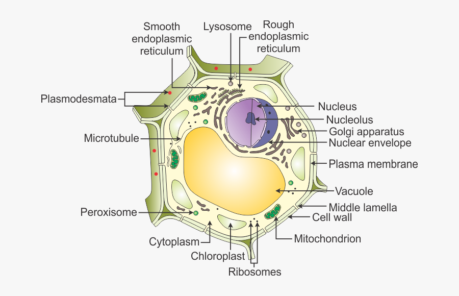
› articlesWhat is Process Mapping? [Definition, Examples & Tools] Aug 18, 2021 · The activities used for process mapping are part of the overall quality management practices of a business. When done correctly, a process map will look at a single objective within a procedure and help measure it against the company's overall goals to ensure that all work being done helps achieve process improvement. quizlet.com › 568033559 › botany-exam-1-chs-1/2/3-4Botany Exam 1 Chs. 1, 2, 3, 4, 5, 6, 7, 12, and 16 Quizzes ... Complete the following diagram by matching each phase of meiosis with the events that transpire during that phase. 1. Chromosomes shorten and thicken for the second time 2. Chromosomes separate and move to opposite poles 3. Chromosomes shorten and thicken for the first time 4. Chromosomes lengthen and cell wall forms making 2 cells 5. PDF Plant Cell Diagram Plant Cell Golgi vesicles Golgi apparatus Ribosome Smooth ER(no ribosomes) Nucleolus Nucleus Rough ER(endoplasmic reticulum) Large central vacuole Amyloplast(star ch grain) Cell wall Cell membrane Chloroplast Vacuole membrane Raphide crystal Mitochondrion Druse crystal Cytoplasm Plant Cell- Definition, Structure, Parts, Functions ... Figure: Labeled diagram of plant cell, created with biorender.com. The typical characteristics that define the plant cell include cellulose, hemicellulose and pectin, plastids which play a major role in photosynthesis and storage of starch, large vacuoles responsible for regulating the cell turgor pressure. They also have a very unique cell division process whereby there is the formation of a ...
Label animal cell - Teaching resources - Wordwall Label Plant and Animal Cell Labelled diagram. by Eawilson. Label Plant and Animal Cell Labelled diagram. by Sciencegeek. Plant and Animal Cell to Label Labelled diagram. by Sstichter. G10 Biology. Plant Cell - Label Organelles Labelled diagram. by Azimmer. Jejunum Histology Slide with Labeled Diagram and ... Normal jejunum wall histology with diagram. If you want to learn the detailed histological features of the jejunum, you may continue this part of the article. I will describe the single layer from the normal jejunum wall histology slide with a labeled diagram. 03 Label the Cell Diagram - Quizlet Start studying 03 Label the Cell. Learn vocabulary, terms, and more with flashcards, games, and other study tools. Plant and Animal Cell: Labeled Diagram, Structure ... Cell Wall: 1. Non-living, rigid, outer boundary. 2. Made up of cellulose, hemicellulose, pectin, lignin, etc. 3. There are many layers, like the middle layer, primary cell wall in a typical plant cell wall. 4. Fungal cell wall is made up of chitin (not cellulose). 5. Protective and provide shape and size. 6. Found only in plant cells. Plasma ...
PPT - What are cell parts and their functions? PowerPoint Presentation - ID:232186
Prokaryotic cell to label - Labelled diagram - Wordwall Prokaryotic cell to label - Labelled diagram nucleoid region, pili, ribosomes, flagellum, plasmid, cytoplasm, plasma membrane, cell wall, capsule. Prokaryotic cell to label Share by Susanmgillen KS5 Biology Like Edit Content More Leaderboard Log in required Theme Log in required Options Switch template Interactives

parts of a cell .pdf - Organelle Function(Job Cell wall The cell wall surrounds the plasma ...
Spirogyra Labelled Diagram Spirogyra Labelled Diagram Biological drawing showing Spirogyra, Single Cell, Biology Teaching Resources by D G Mackean. Draw a neat diagram of Spirogyra and label the following parts: i. Outermost layer of the cell. ii. Organelle that performs the function of.

Cell Wall CH#4 (1st Year Biology) | Cell wall, Environmental research, Biology
Microbiology Test 2 (Chapter 4: Prokaryotes and Eukaryotes ... *Drag each of the labels onto the diagram to correctly label the indicated structures. ... In the figure, which diagram of a cell wall possesses lipid A/endotoxin responsible for symptoms associated with infection? B. In the figure, which diagram of a cell wall has a structure that protects against osmotic lysis? ...

In The Figure Which Diagram Of A Cell Wall Has A Structure That Protects Against Osmotic Lysis ...
Plant Cell Diagram - Science Trends A plant cell diagram, like the one above, shows each part of the plant cell including the chloroplast, cell wall, plasma membrane, nucleus, mitochondria, ribosomes, etc. A plant cell diagram is a great way to learn the different components of the cell for your upcoming exam. Plants are able to do something animals can't: photosynthesize.

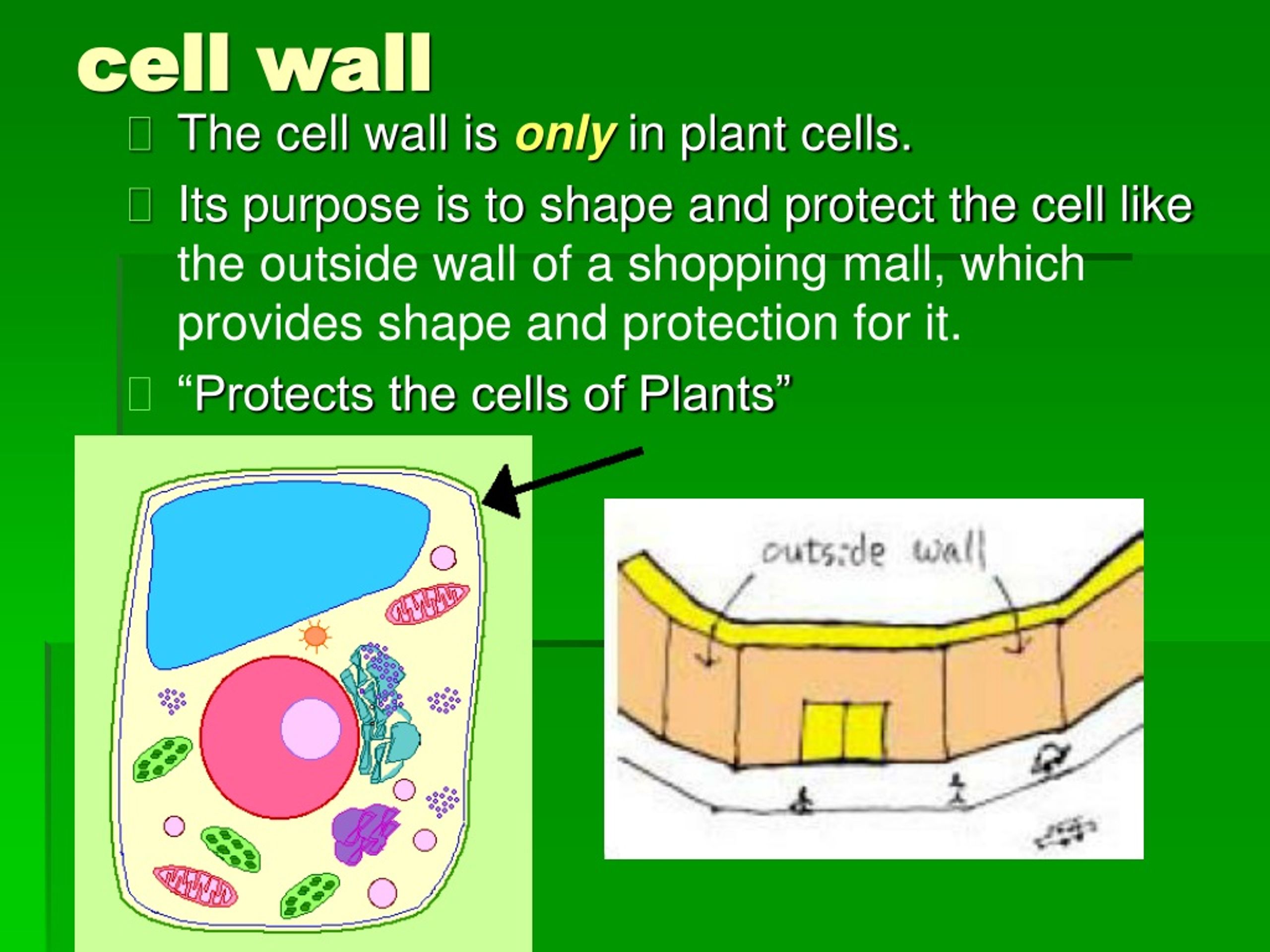
The Plant Cell Is Like A Home..
Plant Cell: Diagram, Types and Functions - Embibe Exams Plant Cell Wall It is a rigid layer that is composed of cellulose, glycoproteins, lignin, pectin and hemicellulose. It is located outside the cell membrane and is completely permeable. The primary function of a plant cell wall is to protect the cell against mechanical stress and to provide a definite form and structure to the cell.

Structure of the Generalized Cell
Meiosis In Plant Cell Diagram Labeled : Functions and Diagram Diagram for Meiosis Meiosis is a type of cell division in which a single cell undergoes division twice to produce four haploid daughter cells. One of the foremost intricate tasks that wellbeing and fitness experts face across their interplay with patients helps them recognise the issues and a way to motivate them in regards to the diagnosis and ...
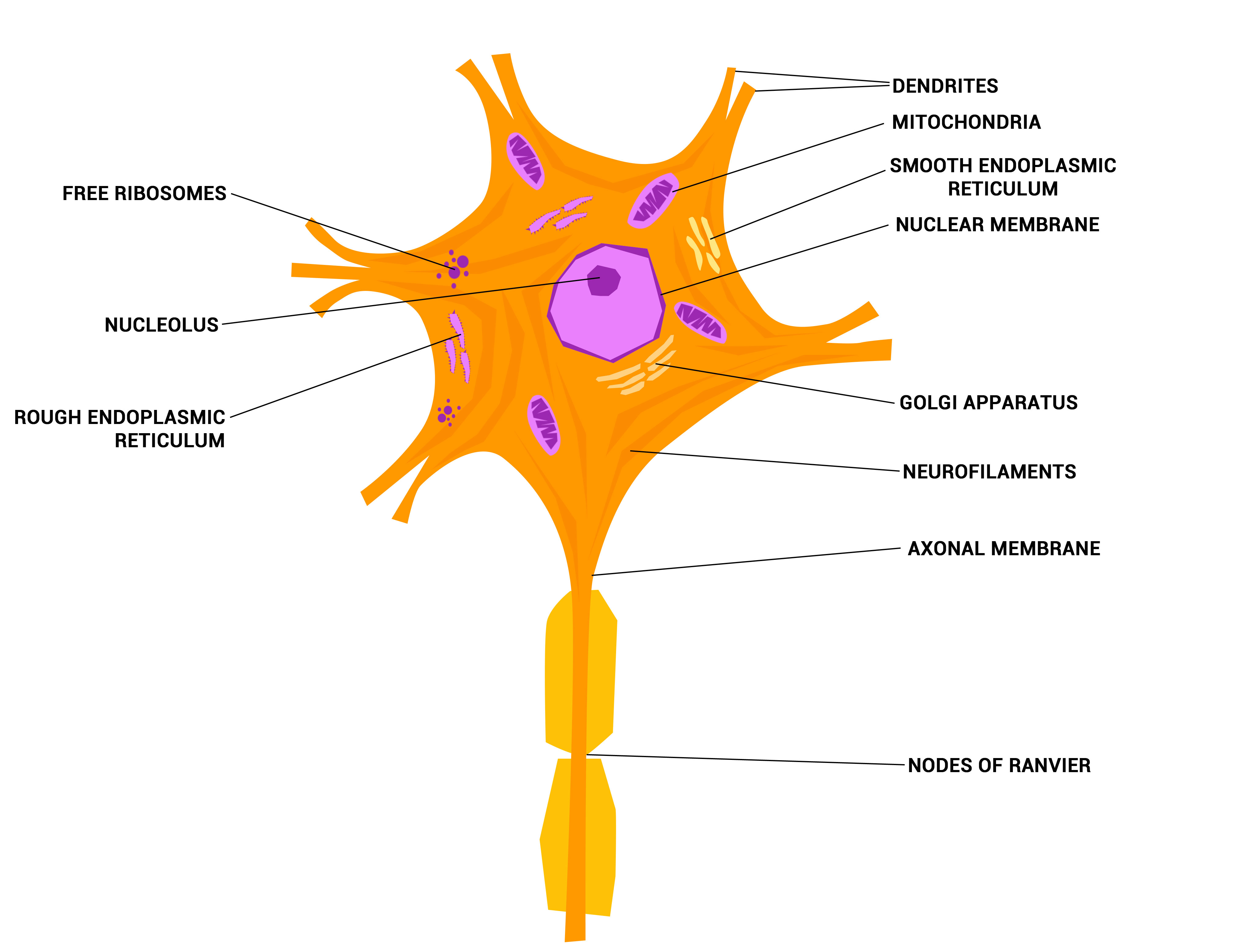
Neurons - The crazy wires in our body. | Doc Jana
PDF Human Cell Diagram, Parts, Pictures, Structure and Functions Diagram of the human cell illustrating the different parts of the cell. Cell Membrane The cell membraneis the outer coating of the cell and contains the cytoplasm, substances within it and the organelle. It is a double-layered membrane composed of proteins and lipids.

Chart Showing Plant Cell Components Stock Illustration - Download Image Now - iStock
Onion Epidermal Cell Labeled Diagram - schematron.org The epidermal cells of onions provide a protective layer against viruses and fungi that may harm the sensitive tissues. Because of their simple structure and.onion epidermal cell labeled onion cell diagram Wide collections of all kinds of labels pictures online. Make your work easier by using a label. Happy Labeling!

Root Hair Cell - Biology
Label Plant and Animal Cell - Labelled diagram - Wordwall Prevuci oznake na odgovarajuće mesto na slici.. Animal Cell, Nucleus, Plant Cell, Cytoplasm, Cell membrane, Mitochondria, Vacuole, Chloroplast, Cell Wall.

Cell Wall Tutorials, Quizzes, and Help | Sophia Learning
qualifications.pearson.com › content › damAS Biology A (Salters-Nuffield) - Edexcel (ii) The student also compared the thickness of the aorta wall of this heart with the thickness of the aorta wall in a giraffe. The thickness of the aorta wall in this heart is 3 mm and in a giraffe it is 15 mm. Give one reason why the aorta wall in a giraffe is much thicker. (1)

Lecture Notes No. 1 for Biology of Plants
Bacteria shapes, structure and diagram - Jotscroll The bacteria shapes, structure, and labeled diagrams are discussed below. Sizes The sizes of bacteria cells that can infect human beings range from 0.1 to 10 micrometers. Some larger types of bacteria such as the rickettsias, mycoplasmas, and chlamydias have similar sizes as the largest types of viruses, the poxviruses.
A Draw A Neat Diagram Of A Plant Cell And Label The - Diagram Of Plant Cell With Labelling ...
Plant Cells: Labelled Diagram, Definitions, and Structure The cell wall is made of cellulose and lignin, which are strong and tough compounds. Plant Cells Labelled Plastids and Chloroplasts Plants make their own food through photosynthesis. Plant cells have plastids, which animal cells don't. Plastids are organelles used to make and store needed compounds. Chloroplasts are the most important of plastids.
Tattoo Blog
PDF PLANT CELL DIAGRAM - abcteach.com for the cell. K. The "control center" of the cell; this contains the cell's DNA. L. A thick, stiff membrane that surrounds the plant cell and supports the plant structure. M. A thin, semi-permeable membrane that surrounds the cell, inside the cell wall. N. An organelle that stores molecules such as starch and pigment. __ 1. Nucleus __ 2.

GCSE biology: Animal and Plant Cells
PDF Plant Cells - Definition, Diagram, Structure & Function Plant Cells - Definition, Diagram, Structure & Function The cell is the basic unit of life in all organisms. Like humans and animals, plants are also composed of several cells. The plant cell is surrounded by a cell wall which is involved in providing shape to the plant cell. Apart from the cell wall, there are other organelles that are

Finley Period 7: December 2010
Cell Organelles- Definition, Structure, Functions, Diagram In a plant cell, the cell wall is made up of cellulose, hemicellulose, and proteins while in a fungal cell, it is composed of chitin. A cell wall is multilayered with a middle lamina, a primary cell wall, and a secondary cell wall. The middle lamina contains polysaccharides that provide adhesion and allow binding of the cells to one another.

Margaret Nielsen: Finished Cell Diagrams with Labels
Animal Cell Labelling Activity | Primary Resources | Twinkl If your students find the Animal Cell Labelling Worksheet useful, this Plant Cell Diagram is a similar labelling activity for plant cells. For something a little more challenging, this Parts of a Cell Cut and Stick Worksheet also challenges students to recall the different components of both animal and plant cells.

diagram of cell wall - Brainly.in
PDF Plant Anatomy: Images and diagrams to explain concepts The cell wall is initially deposited on the surface of the middle lamella. This primary cell wall occurs on the surface of all plant cells. It is substantially composed of cellulose molecules bundled together to form fibrils. The primary cell wall is the only cell wall present in some cells. In other cells a secondary cell wall is deposited inside

The Plant Cell
Cell Wall - Definition, Cell Wall Function, Cell Wall Layers A cell wall is defined as the non-living component, covering the outmost layer of a cell. Its composition varies according to the organism and is permeable in nature. The cell wall separates the interior contents of the cell from the exterior environment. It also provides shape, support, and protection to the cell and its organelles.
Post a Comment for "43 cell wall diagram with labels"